Home » Without Label » Left Hip Muscles Anatomy : Pin On Ortho Bionomy - This anatomical atlas was especially designed for a specific public (radiologists, surgeons, rheumatologists and physicians specializing in musculoskeletal imaging).
Left Hip Muscles Anatomy : Pin On Ortho Bionomy - This anatomical atlas was especially designed for a specific public (radiologists, surgeons, rheumatologists and physicians specializing in musculoskeletal imaging).
Left Hip Muscles Anatomy : Pin On Ortho Bionomy - This anatomical atlas was especially designed for a specific public (radiologists, surgeons, rheumatologists and physicians specializing in musculoskeletal imaging).. Related online courses on physioplus. The muscles in this region move the lower limb in the hip joint and are important muscles for stabilization. Learning the anatomy of your hip will better enable you to pinpoint your pain and work with your doctor to keep it from limiting your life. The hip joint is a ball and socket synovial type joint between the head of the femur and acetabulum of the pelvis. This muscle assists with the external rotation of the hip.
A bursa that sometimes causes problems in the hip is sandwiched between the bump on the outer hip (the greater trochanter) and the muscles and tendons that cross over the bump. Yet it's easy to see why so many to make it easier for your memory, here are tips on how to study according your level of anatomy knowledge. The cavity of the acetabulum the external obturator muscle is short external rotator muscle of hip joint. 3 months later i got acute excrutiating pain in inguinal area. Thigh magnetic resonance imaging the thigh has some of the body's largest muscles.
Illustration Of Left Hip Anterior View Showing Ateries Lateral Femoral Circumflex Arteries Vein And Muscles Orthopaedic Surgical Anatomy Teaching Collection Usc Libraries Digital Collections from digitallibrary.usc.edu We study anatomy at the practical anatomy class we study the human body. Your email address will not be published. Let the left knee fall outward as much as possible. Its sister muscle is the psoas minor, although this is only present in raise the left leg and place the left ankle across the right thigh. This muscle assists with the external rotation of the hip. The muscular system consists of the skeletal muscles and their associated structures. How many of the 11 muscles involved in hip flexion can you name from memory? When the hip muscles are left more or less intact, they are able to support the new hip and shorten recovery times.
4, obturator internus m this webpage presents the anatomical structures found on thigh mri.
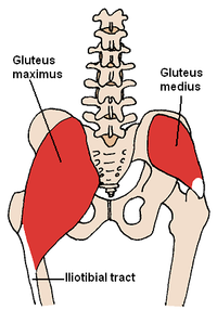
How many of the 11 muscles involved in hip flexion can you name from memory? Keep your left hip extended and your left leg straight. Major lower body muscle groups include leg and hip muscles, largest muscle groups in your body. The hip muscles encompass many muscles of the hip and thigh whose main function is to act on the thigh at the hip joint and stabilize the pelvis. Anatomical terms allow us to describe the body and body motions more precisely. When the hip muscles are left more or less intact, they are able to support the new hip and shorten recovery times. The different anatomical areas of the gluteal region: Pick which works for you and then. Pelvis and acetabulum, with muscle attachment sites. Understanding the anatomy of the lower body, particularly the muscle locations and their functions, will help you to get the most from the exercises and programs presented on this website. 4, obturator internus m this webpage presents the anatomical structures found on thigh mri. Almost all muscles cross at least one joint (moveable connection between two bones) and cause an action across that joint. Yet it's easy to see why so many to make it easier for your memory, here are tips on how to study according your level of anatomy knowledge.
Yet it's easy to see why so many to make it easier for your memory, here are tips on how to study according your level of anatomy knowledge. Gently lower your left leg on the floor. Your email address will not be published. Several muscles cross the front of the hip and create hip flexion, pulling the thigh and trunk toward each other, but probably the most important is the iliopsoas. 4, obturator internus m this webpage presents the anatomical structures found on thigh mri.
Tendinitis And Bursitis Treatment Cincinnati Tendinitis Dayton Oh from www.beaconortho.com Leave a reply cancel reply. Anatomy of the muscular system. The psoas major muscle (usually shortened to just the psoas muscle) is one of the muscles of the posterior abdominal wall and lies not in the retroperitoneum but posterior to it, in the iliopsoas compartment. Thigh magnetic resonance imaging the thigh has some of the body's largest muscles. Related online courses on physioplus. The different anatomical areas of the gluteal region: Its sister muscle is the psoas minor, although this is only present in raise the left leg and place the left ankle across the right thigh. Leave a comment cancel reply.
Raise your left leg as high as possible.
When the hip muscles are left more or less intact, they are able to support the new hip and shorten recovery times. If left unstretched, shortened hip flexors affect the position of the pelvis, which in turn affects the position and movement of the lower back. In human anatomy, the muscles of the hip joint are those muscles that cause movement in the hip. The different anatomical areas of the gluteal region: Leave a reply cancel reply. It is a flat, triangular muscle on the anterior wall of the pelvis. Microscopic anatomy of skeletal muscle. We study anatomy at the practical anatomy class we study the human body. Anatomy 3d atlas allows you to study human anatomy in an easy and interactive way. Anatomical terms allow us to describe the body and body motions more precisely. There are a lot of muscles of the hip and thigh. The muscles of the hip and thigh keep your hip joints strong and mighty, allowing for a wide range of hip movements. I pulled some muscles on left hip hiking.
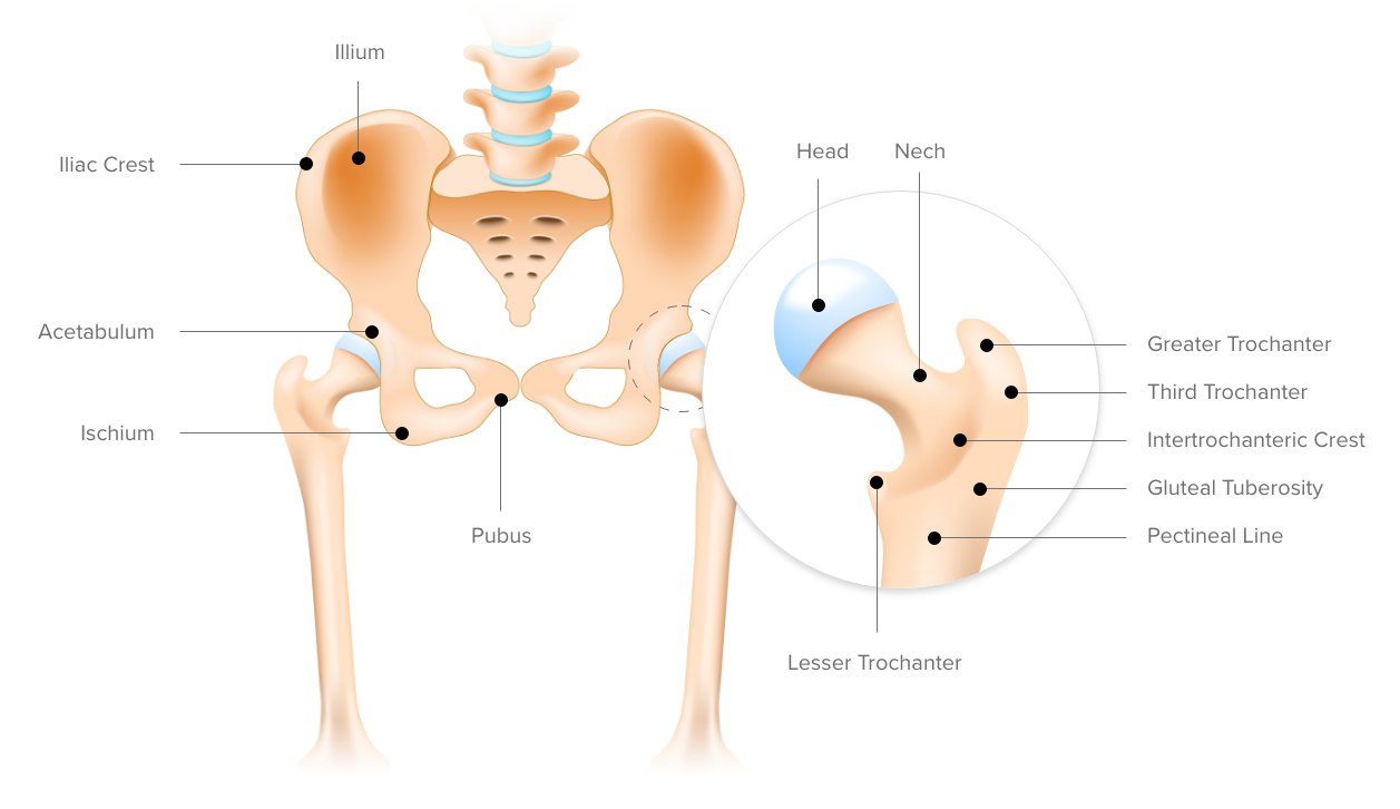
for detailed anatomy of pelvic bones, read anatomy of hip bone. The psoas major muscle (usually shortened to just the psoas muscle) is one of the muscles of the posterior abdominal wall and lies not in the retroperitoneum but posterior to it, in the iliopsoas compartment. This anatomical atlas was especially designed for a specific public (radiologists, surgeons, rheumatologists and physicians specializing in musculoskeletal imaging). The muscles and the bones are under the layer of subcutaneous fat. If left unstretched, shortened hip flexors affect the position of the pelvis, which in turn affects the position and movement of the lower back.
Muscles Of The Hip Wikipedia from upload.wikimedia.org 3 months later i got acute excrutiating pain in inguinal area. If left unstretched, shortened hip flexors affect the position of the pelvis, which in turn affects the position and movement of the lower back. A bursa that sometimes causes problems in the hip is sandwiched between the bump on the outer hip (the greater trochanter) and the muscles and tendons that cross over the bump. Muscle movements, types, and names. Major lower body muscle groups include leg and hip muscles, largest muscle groups in your body. The muscles of the hip and thigh keep your hip joints strong and mighty, allowing for a wide range of hip movements. Your email address will not be published. It's hard to remember them all!
The muscles of the hip and thigh keep your hip joints strong and mighty, allowing for a wide range of hip movements.
How frequently and with what power these electric stimuli. Learning the anatomy of your hip will better enable you to pinpoint your pain and work with your doctor to keep it from limiting your life. Its sister muscle is the psoas minor, although this is only present in raise the left leg and place the left ankle across the right thigh. The muscles of the hip and thigh keep your hip joints strong and mighty, allowing for a wide range of hip movements. We study anatomy at the practical anatomy class we study the human body. Several muscles cross the front of the hip and create hip flexion, pulling the thigh and trunk toward each other, but probably the most important is the iliopsoas. Anatomy 3d atlas allows you to study human anatomy in an easy and interactive way. This anatomical atlas was especially designed for a specific public (radiologists, surgeons, rheumatologists and physicians specializing in musculoskeletal imaging). There are a lot of muscles of the hip and thigh. Pelvis and acetabulum, with muscle attachment sites. A bursa that sometimes causes problems in the hip is sandwiched between the bump on the outer hip (the greater trochanter) and the muscles and tendons that cross over the bump. How many of the 11 muscles involved in hip flexion can you name from memory? Microscopic anatomy of skeletal muscle.